The Mysterious Episodes of Mary: A Case Study on Neuroanatomy
Episode 1
Mary Lazarro, a 44-year-old mother of two, made an appointment with her physician after experiencing a prolonged episode of numbness in her chin and lower lip. Two days prior to her appointment, she felt a prickling sensation like "pins and needles" at the right corner of her mouth. The sensation extended to her lower lip and chin. The examination revealed only a superficial hypoesthesia of the chin and lower lip (numb chin syndrome). There was no clinical evidence of palpable regional lymph nodes or other systemic or neurologic abnormalities. Her physician scheduled her for a CT scan of the affected region. These tests showed no abnormalities in the jaw, neck, or pharynx. The numbness and hypoesthesia spontaneously disappeared gradually over a few weeks.
Episode 2
Four months later, while eating dinner with her family, Mary felt a stabbing pain in her upper jaw and teeth that radiated out to the side of her nose. Over the next several days, she experienced several more episodes of this intense pain. A visit to the dentist revealed no abnormalities and she was referred to her physician for an evaluation. Prior to her appointment, she noticed that the symptoms were subsiding as they had previously. Her physician scheduled an appointment for a complete neurological exam the following week.
Episode 3
Three nights prior to her scheduled visit to the neurologist, Mary stopped at an intersection and experienced intense double vision when looking to the right to check for traffic. The double vision was less intense when looking forward, and her vision when looking left was unaffected. Her husband noticed that her right eye appeared to be turned slightly inward when she looked straight ahead. A day later, Mary noticed that the vision in her left eye started to blur. The neurologist later suggested that the two visual problems she was experiencing were related. The double vision when looking right was found to be caused by cranial nerve palsy-a form of muscle paralysis caused by a dysfunction in one of the cranial nerves. The problem with the left eye was diagnosed as optic neuritis (inflammation). Both of these signs and symptoms, along with the previous episodes, pointed to a diagnosis of multiple sclerosis (MS). The neurologist prescribed oral steroids and ordered an MRI. As with her previous episodes, Mary's visual symptoms began to diminish over time.
Finale
The results of the MRI, shown below, were consistent with a diagnosis of relapsing-remitting multiple sclerosis (RRMS). Relapsing-remitting multiple sclerosis is a form of MS in which symptoms randomly flare up (Mary's episodes) and then resolve on their own. The lesions seen on the MRI on the left were associated with another episode in which Mary experienced sensory and motor disjunction in her left lower extremity. A subsequent MRI (image on the right) appeared to show improvement after three months.
Short answer questions
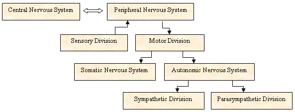
1. Related to Episode 1: Using the flowchart below, identify the part of the human nervous system that is usually associated with symptoms of hypoesthesia and paresthesia.

2. Related to Episode 1: Which of Mary's cranial nerves is affected in this episode?
3. Related to Episode 2: Which of Mary's cranial nerves is affected in this episode?
4. Related to Episode 3: Name all of the cranial nerves that are involved with eye movements.
5. Related to Episode 3: Which of Mary's affected cranial nerves is responsible for her double vision when looking right? Why does she not experience double vision when looking left?
6. Related to Episode 3: Which of Mary's affected cranial nerves is responsible for her blurred vision?
7. Related to Finale: In the MRI images shown in the case, you can see the lesions as bright "white spots" on the brain. Using what you know about the structure of a neuron, explain what is causing this spot to appear in the MRI.
8. Related to Finale: Three months later, you can see that the spots in the MRI appear to be smaller. Using what you know about the structure of a neuron, explain what is happening to the neurons in the area where the lesions are disappearing.